
Description
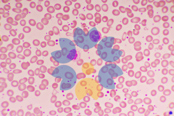
This is a microscopic image of a blood smear stained with a dye, likely Wright-Giemsa stain, which is commonly used in hematology to differentiate blood cells.
Observations:
- Red Blood Cells (RBCs): The numerous round, pink-stained cells in the background are erythrocytes. They appear mostly uniform in size and shape, with a pale center, indicating the presence of normal biconcave disc-shaped RBCs.
- White Blood Cells (WBCs): There are a few larger, dark purple-stained cells, which are leukocytes (white blood cells). These could be neutrophils or lymphocytes, responsible for immune response.
- Platelets: Small, purple-stained fragments are visible, which are platelets. These are involved in blood clotting.
Possible Interpretation:
This appears to be a normal peripheral blood smear, but variations in RBC shape, size, or WBC count could indicate underlying conditions like anemia, infection, or hematological disorders. A detailed clinical correlation would be required for an accurate diagnosis.

Reviews
There are no reviews yet.